Learn Emphysema: Diagrams


View Enlarged Diagram
Print Diagram
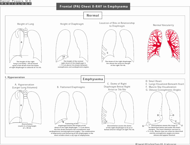
Frontal (PA) Chest X-RAY in Emphysema
Emphysema can be inferred on chest x-rays by various findings, some of which can be measured. This diagram shows some of the findings associated with emphysema that makes one suspicious on a frontal (PA) chest radiograph that the patient has underlying emphysema. Hyper or overinflation is the most reliable finding. The diagnosis has to be proven, however, on the basis of the patient's history, clinical examination, pulmonary function testing, and if necessary CT or HRCT. There are some individuals that have an elongated body frame, who are malnourished, who are very athletic with very full and elongated lungs, such as in runners, or who have other disease processes including asthma that can mimic emphysema.Lateral Chest X-Ray Changes in Emphysema
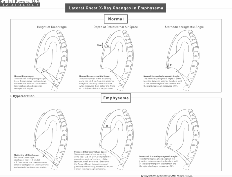
The normal and abnormal findings on the lateral plain radiograph are documented, some of which can be measured, if so desired.Frontal (PA) Chest X-Ray Changes in Emphysema
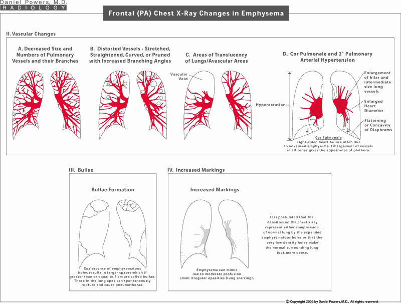
Multiple other changes can be identified on chest x-rays including changes to the vasculature such as decreasing size and numbers of pulmonary vessels and their branches, distorted vessels with increasing branching angles, and areas of translucency or avascular regions within the lungs. In end-stage disease, right-sided heart failure can occur from cor pulmonale with associated secondary pulmonary arterial hypertension. Other findings associated with emphysema occasionally seen on chest x-ray include large holes known as bullae or increased, what appear to be, interstitial markings.


