Learn Silicosis: Diagrams
View Enlarged Diagram
Print Diagram
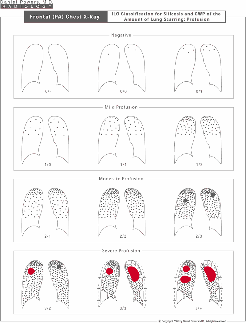
Frontal (PA) Chest X-Ray; ILO Classification for Silicosis and the Amount of Lung Scarring
1silicosis-ilo-profusion-classification
This uses a 12-point system, in which there are 4 general categories of disease appearance - "0" for negative, "1" for mild, "2" for moderate, and "3" for severe disease. Because there can be variations in the amount of disease present, plus or minus categories are included. An example would be a 2/2 profusion is considered a definitive moderate amount of disease process. A 2- would be a 2/1, which means the doctor considered that this was a 2 or moderate amount of disease first with a second consideration that it was slightly less, tending towards a mild amount of disease. A 2+ category would be called a 2/3, meaning that this was definitely a moderate amount of disease as a first consideration, but as a second consideration there was more advanced to severe disease.
ILO Parenchymal Classification for Silicosis
2silicosis-plus-cwp-ilo-classification
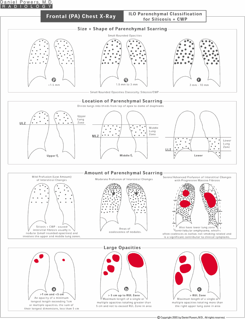
The small rounded opacities are classified in terms of their size - "p", "q" or "r"; their location - whether they are in the upper, middle or lower lung zones; the amount of scarring as mild, moderate or severe which is further related to the 12-point ILO classification system, and whether there are "Large Opacities" - masses that measure greater than 1 cm in diameter.
Sequential Chest X-Ray Parenchymal Changes in Silicosis
6silicosis-plus-cwp-sequential-chest-x-ray-changes
This diagram shows sequential changes that can occur after the inhalation of coal dust and/or free crystalline silica. Compared with asbestosis, these two disease processes are vastly different. Asbestosis tends to have irregular to linear densities in the mid to lower lung zones associated with pleural plaquing or diffuse pleural thickening. Silicosis and CWP tend to have small rounded opacities in the upper to mid-lung zones, which can be associated with central hilar lymph node enlargement having eggshell calcifications and the absence of diffuse pleural thickening or pleural plaquing. However, coalescence of subpleural nodules can mimic plaques and these can develop calcifications and occasionally rounded atelectasis. In addition, these diseases develop progressive massive fibrosis usually involving the upper lung zones, which eventually cause distortion of the intrathoracic contents and are associated with resultant emphysematous changes.
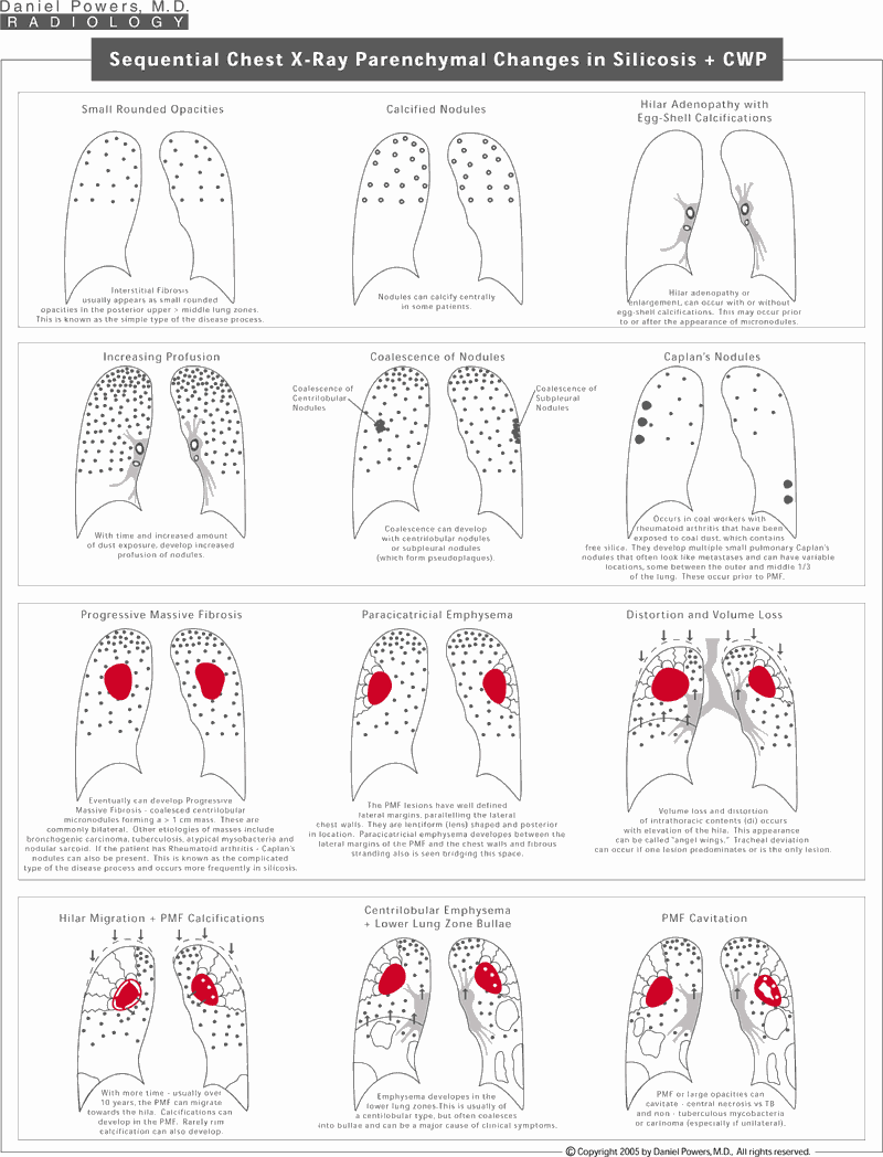
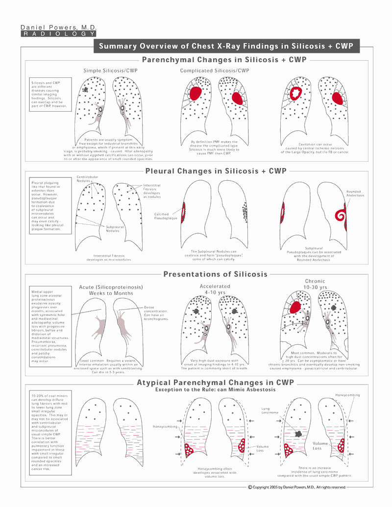
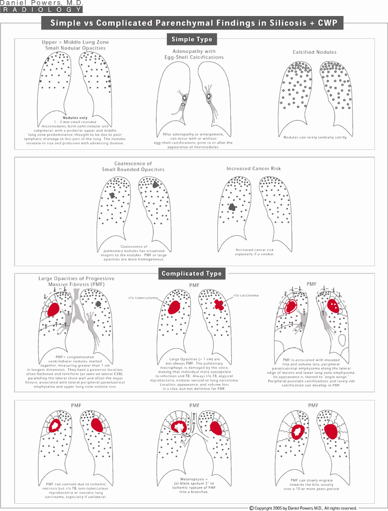
Simple versus Complicated Parenchymal Findings in Silicosis
7silicosis-plus-cwp-simple-plus-complicated-chest-x-ray-findings
The disease process is separated into two general types, the simple type and the complicated type. The main difference being, that the complicated type is a more advanced form of the disease process that is associated with progressive massive fibrosis. Changes that can occur in these two conditions are visually demonstrated.
Summary Overview of Chest X-Ray Findings in Silicosis
8silicosis-plus-cwp-summary-overview-of-chest-x-ray-findings
Parenchymal changes identified include small rounded opacities, coalescence of nodules and large opacities. Pleural changes in silicosis are rare, but coalescence of subpleural nodules can result in pseudoplaques, which can calcify and occasionally be associated with rounded atelectasis. Silicosis unlike CWP can have an additional form of the disease known as acute silicosis or silicoproteinosis. There are 3 basic presentations of silicosis - acute or silicoproteinosis, accelerated and chronic forms. The accelerated and chronic forms have both the simple and complicated types of the disease process, but the accelerated form obviously occurs at a faster pace. Coal Workers' Pneumoconiosis can have an atypical presentation different from silicosis, which mimics asbestosis. This can often be differentiated from asbestosis by the coal workers' occupational history, the absence of pleural plaquing and occasionally by the presence of some small rounded opacities.
© 2024 Daniel Powers, MD   800-222-6768